
Dental Radiology
Dental radiology is the core diagnostic modality of veterinary dentistry. Dental radiographs assist in detecting hidden painful pathology, estimating the severity of dental conditions, assessing treatment options, providing intraoperative guidance, and also serve to monitor success of prior treatments. Unfortunately, most professional veterinary training programs provide little or no training in veterinary dentistry in general or dental radiology in particular.
3D Imaging
3D imaging in dentistry is a valuable tool that allows dentists to create accurate and detailed images of the teeth, jaw, and surrounding tissues. There are various types of 3D imaging technologies used in dentistry, including Cone Beam Computed Tomography (CBCT), digital radiography, and intraoral scanning.
CBCT (Cone Beam Computed Tomography)
CBCT stands for Cone Beam Computed Tomography, which is a medical imaging technology that uses X-rays to produce three-dimensional images of dental structures, such as teeth, jaws, and the surrounding bones. CBCT provides a detailed and accurate view of the oral and maxillofacial region, which can be used for diagnosis, treatment planning, and evaluation of the effectiveness of treatment.
CBCT is commonly used in dental implant planning, orthodontic treatment planning, and evaluation of impacted teeth. It is also useful in the diagnosis of dental and maxillofacial conditions such as jaw tumors, temporomandibular joint disorders, and sinus problems.


2D Imaging
Intraoral radiography involves placing a small film or digital sensor inside the patient’s mouth to capture detailed images of individual teeth and their roots. This type of imaging is often used to diagnose cavities, check the fit of dental restorations, and evaluate the bone levels around teeth.
OPG (Orthopantomogram)
- Wisdom teeth: OPGs can show the position and development of wisdom teeth, which can be helpful in deciding if they need to be removed.
- Tooth decay: OPGs can show areas of decay that are not visible during a visual exam.
- Gum disease: OPGs can show the extent of gum disease and help plan treatment.
- Jaw problems: OPGs can show any jaw abnormalities, such as cysts, tumors, or fractures.
- Dental implants: OPGs can help plan dental implant surgery by showing the thickness and density of the jawbone.
RVG (RadioVisioGraphy)
The RVG system works by using a small electronic sensor that is placed inside the patient’s mouth. The sensor captures an image of the teeth and surrounding tissues by converting the x-ray energy into an electrical signal, which is then sent to a computer for processing. The resulting image is displayed on a computer screen, where it can be analyzed by the dentist or dental radiologist.
The RVG system has several advantages over traditional film-based dental radiography systems. It produces images more quickly, with a lower radiation dose to the patient. Additionally, RVG images can be manipulated and enhanced digitally, allowing the dentist to better diagnose and treat dental problems.

Online Consultation
Get the dental care you need from the comfort of your own home with our convenient and secure online consultation services.
Technology and Equipment
We strive to bring to you the best and world class technologies for excellent results.
Certified
Our highly skilled and trained team will treat you with compassion, kindness, and competence to ensure you have an outstanding experience.
Book An Appointment Now
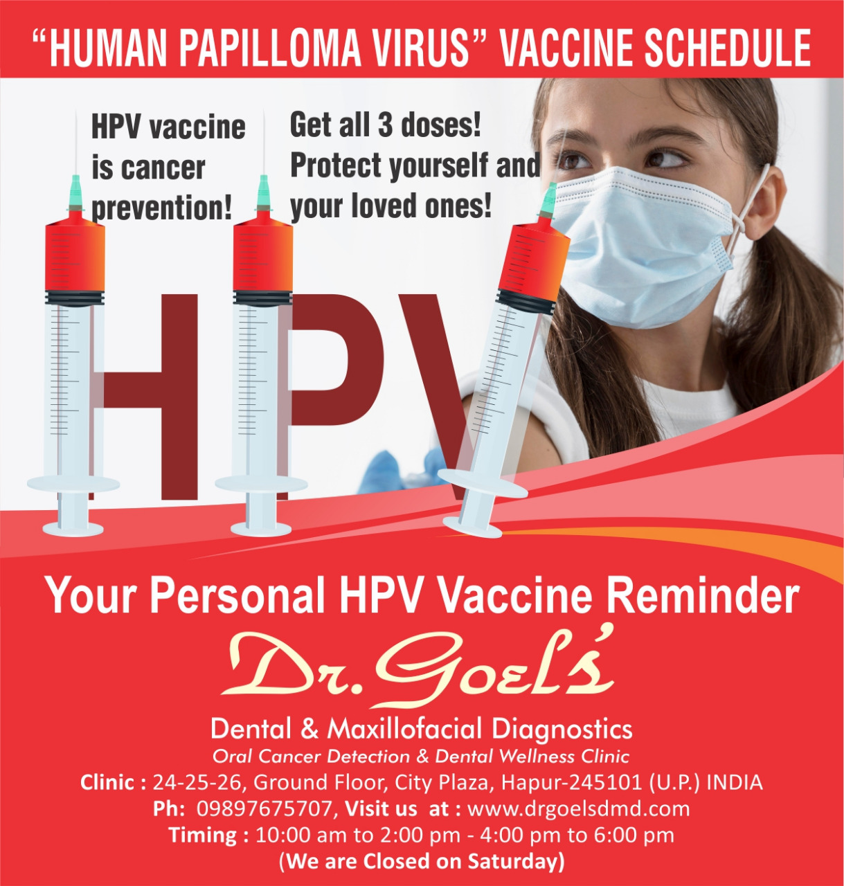
Get your “Oral Cancer Prevention HPV Vaccine” booked today